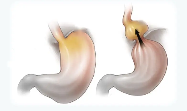
It is located at the end part of the stomach and oesophagus under the muscle bundle separating the chest and abdominal cavity called diaphragm, thus in the abdominal cavity. The upward displacement of the stomach through the hole where the oesophagus pierces the diaphragm is called “Hiatal Hernia”, ie stomach hernia.
The stomach is an organ that produces acid to digest food. The oesophagus is sensitive to acid and is affected by the acid reaching the oesophagus from the stomach during the hernia. Prolonged contact of the oesophagus with acid can cause precancerous lesions called “Barret oesophagus”.
The most prominent symptoms are retrosternal pain, burning and fluid coming into the mouth, which vary in severity with position. In addition, depending on the type and severity of the hernia, symptoms such as early satiety, postprandial pain, feeling of fullness, bitter liquid in the mouth and constipation may be observed.
In endoscopy-guided imaging, oesophageal changes are especially important for us surgeons. In particular, oesophagitis that does not improve despite medical treatment, in other words, the esophagus being affected by acid is the reason for surgery.
In addition, methods such as tomography, barium esophago – gastric radiography and pH manometry are also used in diagnosis.
Hernias unresponsive to medical treatment, herniation of the fundus part of the stomach (Paraesophageal Hernia / Type 2 hernia and Type 3 Hernia), herniation of organs or tissues other than the stomach (Type 4 hernia) cannot be treated without surgery.
Laparoscopically, the stomach is pulled back into the abdominal cavity and the diaphragmatic opening is closed. Depending on the situation, a patch can be placed in the hernia area. The upper region of the stomach is released and rotated 270-360 degrees around the lower end of the oesophagus, and the stomach is fixed to the oesophagus again and prevented from escaping upwards.


